3D Cell Culture Model and Development
The preclinical evaluation of molecules is mainly based on the use of cells in monolayer culture. As this model stays highly artificial regarding cells organization, it can only result in unrepresentative and unpredictive responses.
To better mimic the in vivo biology and close the gap between 2D cell culture and animal models in terms of throughput, cost and relevance, Imactiv-3D offers a 3D cell culture models service.
Our team of biologists has expertise in the manipulation of numerous cell lines and can also develop new protocols for 3D cell models that will best answer our clients' research questions:
Our team of biologists has expertise in the manipulation of numerous cell lines and can also develop new protocols for 3D cell models that will best answer our clients' research questions:
- 30 cell lines available
- Development of spheroids in units or included in matrix
- Custom 3D cell models development: any cell line or matrix
- Quality control of 3D cell models by measuring morphological (diameter, volume, eccentricity, roundness...) and functional (viability, proliferation, differentiation...) criteria
Which cell line do you need?
Cell Viability and Cytotoxicity
The in vitro evaluation of cell viability and cytotoxicity is a key step in the assessment of the biological activity of a new pharmacological compound or a natural product.
To reach this objective, Imactiv-3D offers its expertise in cell culture, cancer cell biology and pharmacology to test your compounds using 2D and 3D cell culture systems.
We can provide two different approaches:
We can provide two different approaches:
- Classical molecular probes (WST1/Alamar blue…) can be combined with a fluorescent plate reader analysis to estimate EC50 or IC50 of your molecule/compound.
- A PI labelling (or other specific labelling of cell death) combined with an automated imaging and cell counting analysis. This can be done together with other specific labelling such as proliferation.
Cell Proliferation Assays
The proliferation rate of in vitro cultures or ex vivo samples is a parameter of interest to characterize the evolution of a model over time or its response to a treatment.
Based on a strong expertise in cell proliferation staining, in 2D and 3D cell models, Imactiv-3D offers services to best answer your questions.
We propose various options to label proliferating cells:
We propose various options to label proliferating cells:
- use of molecular probes such as EdU (exclusively on living samples)
- use of immunofluorescence staining of proteins of interest such as Ki67
3D Immunofluorescence
The detection and visualization of specific cell markers or structures by immunofluorescence while preserving 3D sample morphology is not an easy task.
However, antibody labelling allow to stain a boarder range of target and that in a more specific way than dye labelling.
The 3D immunofluorescence principle is the same than for 2D immunofluorescence with consecutive steps of permeabilization, saturation and antibody incubation. Playing on time and solutions composition able us to adapt the protocol to best suited for 3D samples.
Imactiv-3D has a strong expertise in 3D immunofluorescence on spheroids and works actively to adapt tunable in-house protocol to numerous samples in size and structure such as bioprinted samples or ex vivo organs/tissues. Coupling our in toto antibody labelling method with 3D imaging and 3D analysis, will enable you to characterize and analyze our sample without loss of information.
Migration and Penetration Assays
The study of molecule diffusion is an important issue in pre-clinical study.
It allows for example to follow the infiltration of immune cells into tumors or molecule diffusion into biological tissues.
These types of analysis cannot be done on 2D cell culture and is very costly and time consuming in animal models. The 3D cellular model is the appropriate alternative as it is less expensive than animal models and more relevant than monolayer cultures. Imactiv-3D internal research works actively on this subject to be able at short time to follow antibody penetration into spheroid models. This test/service will be particularly relevant for immunotherapy to follow the fate of a therapeutic antibody in a microtumor-like system.
In parallel, Imactiv-3D is also working at the detection and analysis of immune cells penetration on 3D cellular model within a European consortium. This work is under progress but we already have some promising results. In further applications, Imactiv-3D will also work on transferring these technologies within more complex models such as bioprinted tissues or ex vivo organs.
High Content Screening
In pre-clinical studies, there is a need to test a large number of drug candidates. To address this issue, Imactiv-3D has access to imaging facilities allowing high content screening (HCS).
We can offer several microscopy technologies coupled with automatic plate readers to best fit your needs (wide-field, confocal spinning disk or structured light).
Using the confocal spinning disk, we routinely study monolayer culture of pre-adipocytes and quantify in 3D the lipidic accumulation in lipid droplets or the matrix production with quantification of a density of collagen and fibronectin fibers.
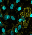
Quantification of lipid droplets in preadipocytes
Read more
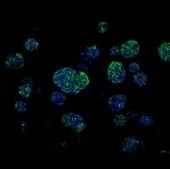
3D Quantification of multispheres growth in various scaffolds
Read more
Using the structured light automated microscope, we have developed a procedure to test compounds on 3D multispheres models embedded in matrix.
A robust statistical analysis can be performed on the multispheres size from an image stack of about 200µm depth.
Infection
Imactiv-3D developed a strategic alliance with NeoVirTech that aims to develop a unique platform with both expertise and innovative tools to move virology projects forward with robust data.
- Preclinical evaluation of compounds efficacy on innovative 3D models by monitoring viral infection and replication using state-of-the-art live imaging techniques. A collection of around 30 viruses is available.
- 3D quantification and visualization of infection (spheroids, organoids, tissues, organs…) combined with staining of a marker of interest. 3D reconstruction for marketing and communication purposes.
Adipocyte Biology
Thanks to our close partnership with D.I.V.A. Expertise, a CRO specialized in research on human adipose tissue, Imactiv-3D is able to provide new services to deepen the exploration in adipocyte biology, for example:
Intra-cellular analysis of lipid accumulation:
- Differentiated cells percentage
- Total intra-cytoplasmic lipid accumulation
- Size and number of lipid droplets
- Imaging (confocal – structured light microscopy)
- Segmentation: fibers search algorithm developed in-house for quantification
- Skeletonization: Fibers length and thickness
- Output: illustration and dataset (fibers length, thickness and quantity)
- Automated UCP-1 quantification