-
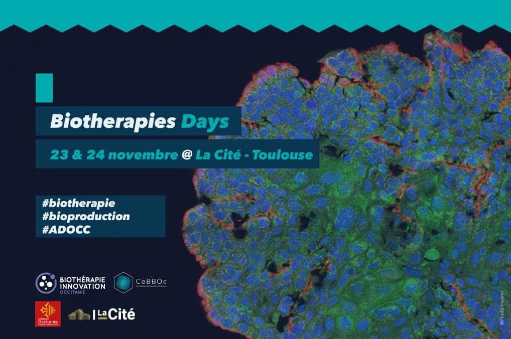
Biothérapies Days
23 nov 2023 -
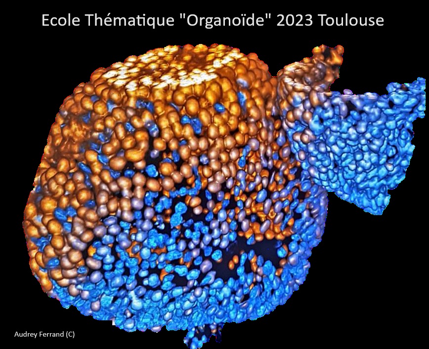
GDR Organoïde
9 oct 2023 -
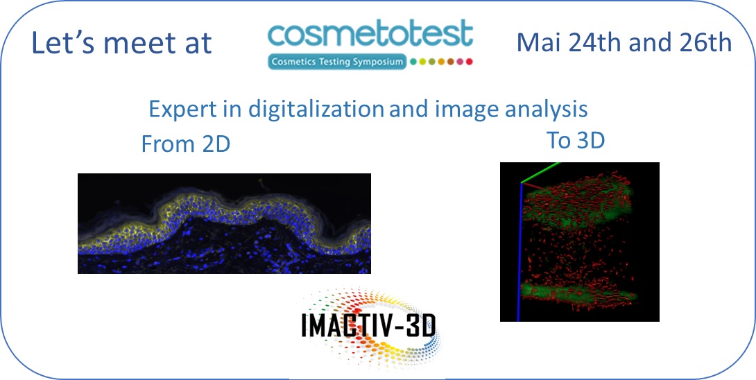
Cosmetotest
23 may 2023 -
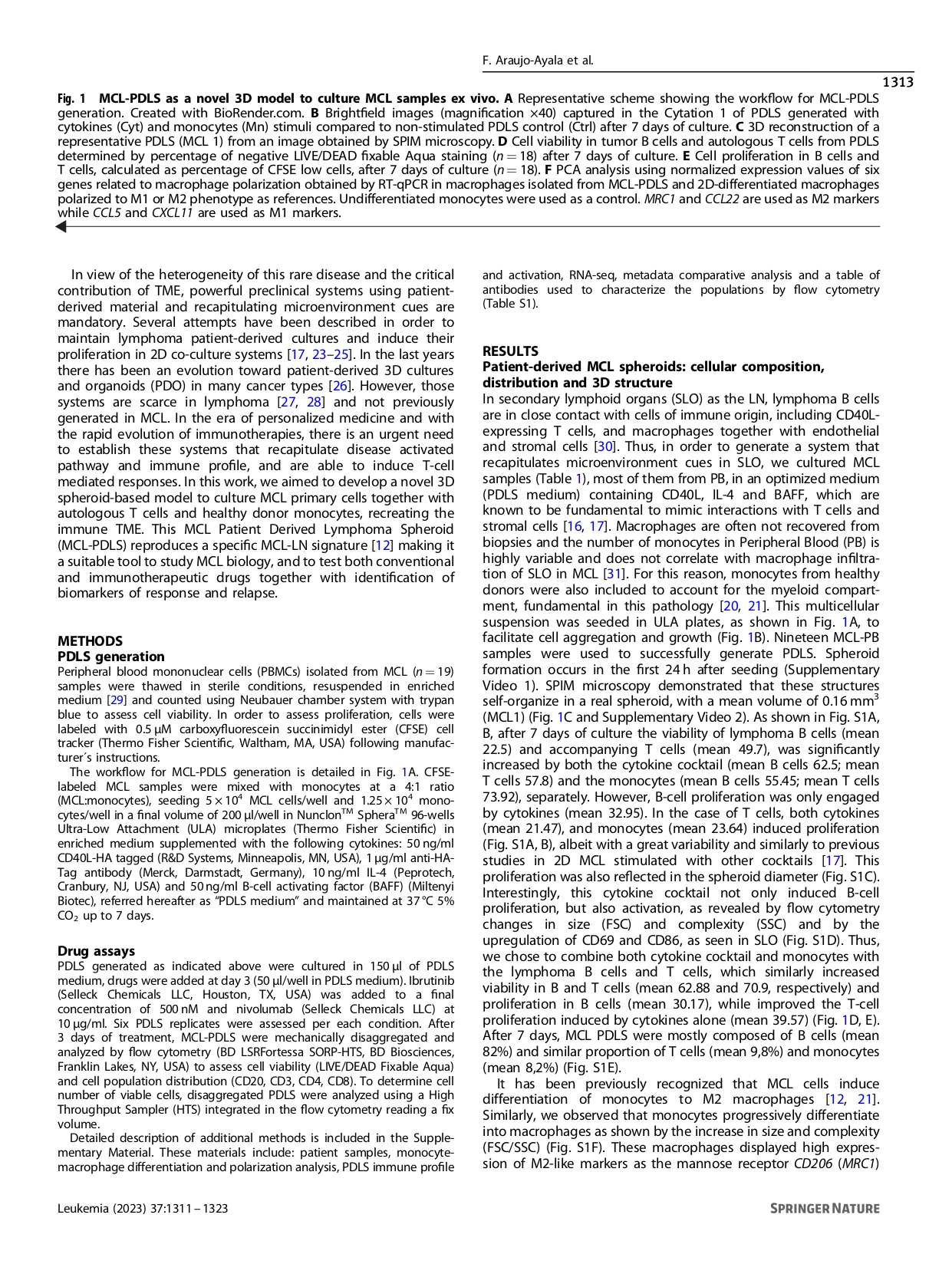
Article
7 apr 2023 -
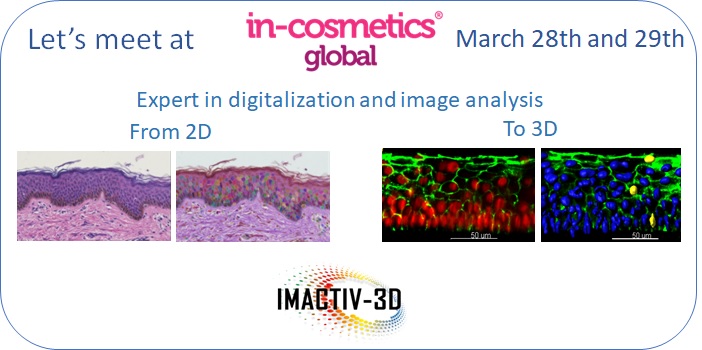
In-Cosmetics Global
28 mar 2023 -
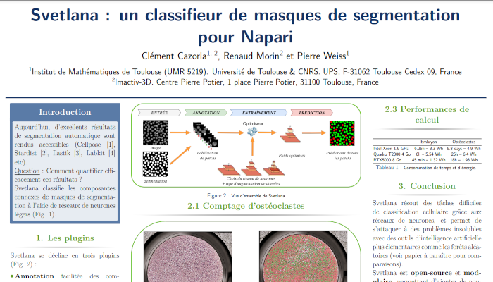
Svetlana, le classifieur de masques de segmentation pour Napari
29 nov 2022 -
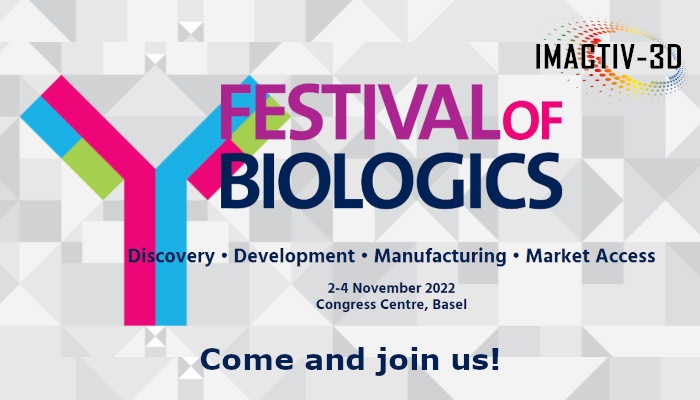
Festival of Biologics 2022
2 nov 2022 -
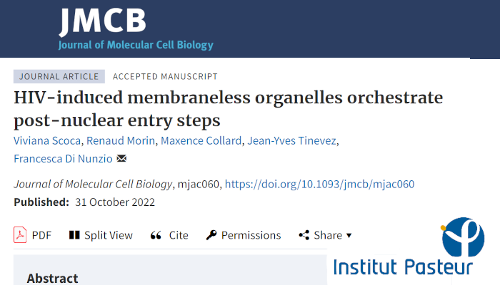
Scientific paper with Institut Pasteur on HIV
31 oct 2022 -
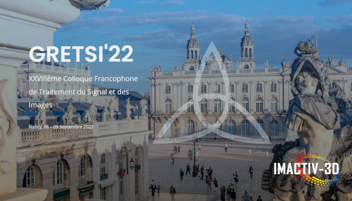
GRETSI'22 Symposium on image and signal processing
3 sep 2022 -
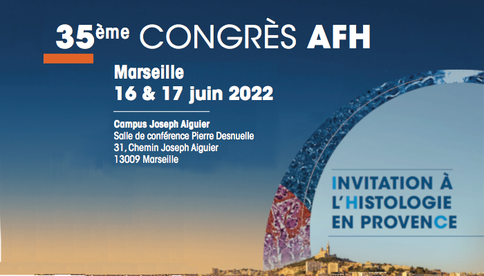
35ème congrès AFH - Histologie
16 jun 2022 -
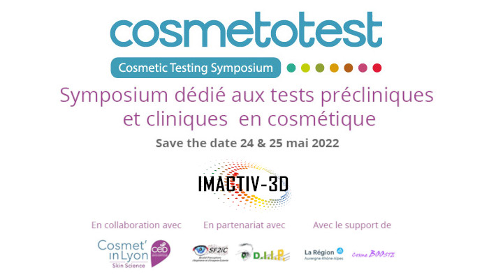
Cosmetotest - Preclinical and clinical testing in dermocosmetics
24 may 2022 -
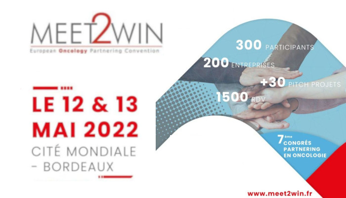
Meet2Win - Oncology Partnering Convention 2022
12 may 2022 -

Scientific paper on Neuro-muscular junctions quantification
19 apr 2022 -

Meet us at In-Cosmetic Global
5 apr 2022 -
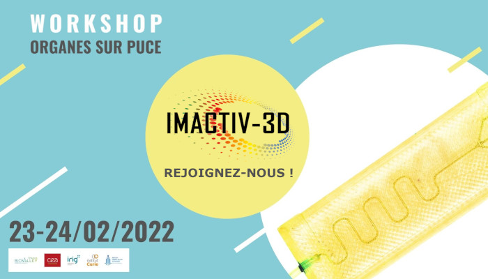
Workshop Organ on Chips
23 feb 2022 -
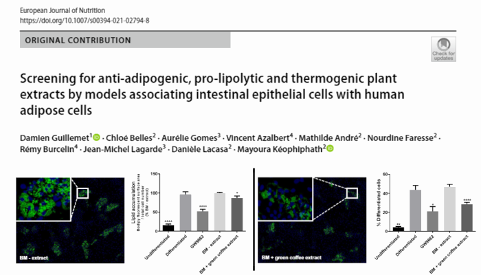
New scientific paper on lipid accumulation
1 feb 2022 -
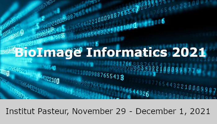
Bioimage Informatics conference organized by Institut Pasteur
1 dec 2021 -
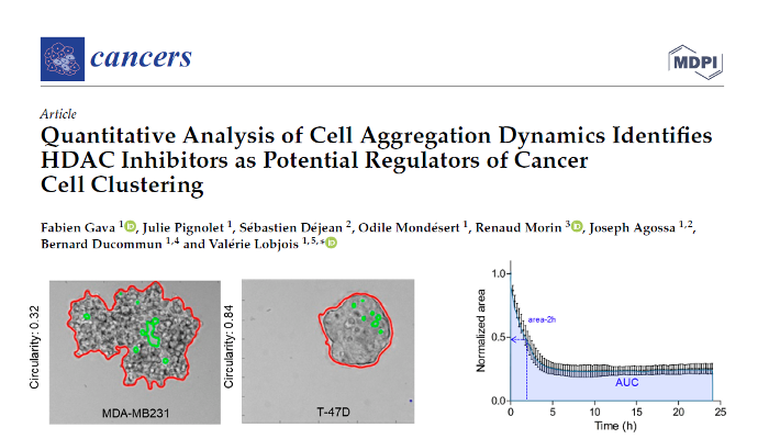
Cancer cell aggregation article published in Cancers
20 nov 2021 -
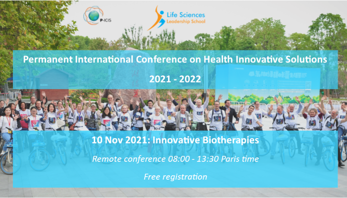
International Conference on Health Innovative Solutions
10 nov 2021 -
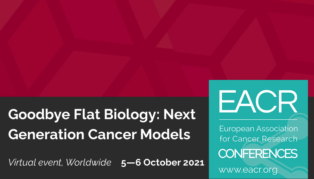
EACR conference: Goodbye Flat Biology, Next-generation Cancer Models
5 oct 2021 -
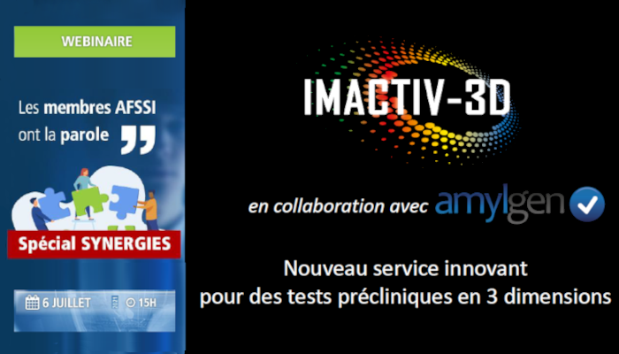
Webinar membre AFSSI : Amylgen & Imactiv-3D
5 jul 2021 -
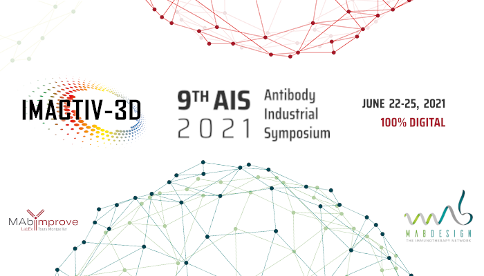
Imactiv-3D at the Antibody Industrial Symposium 2021
21 jun 2021 -
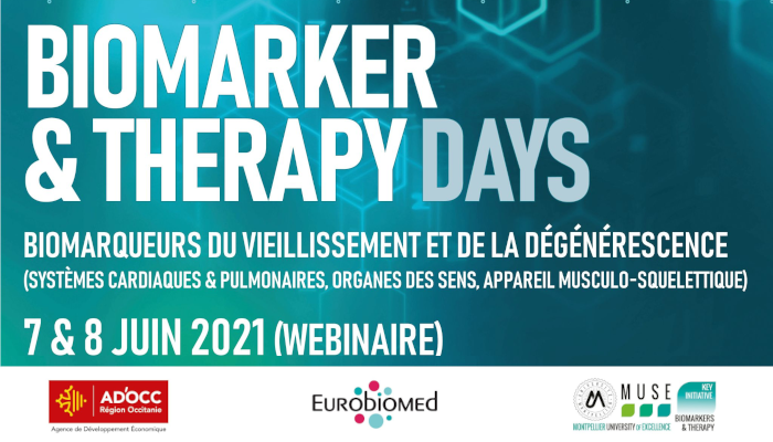
Biomarker and Therapy Days: Aging and Degeneration
7 jun 2021 -
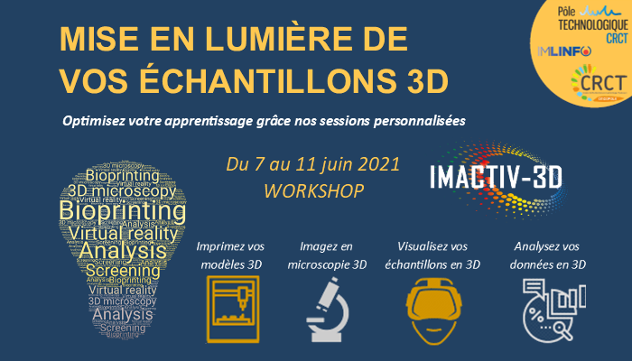
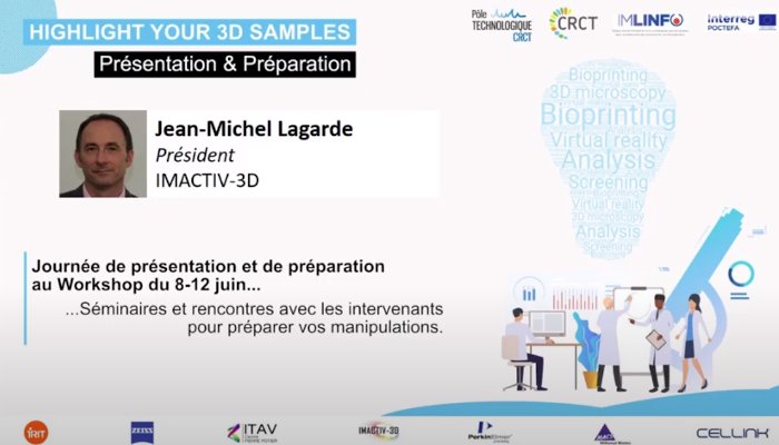
Workshop "Highlight your 3D samples"
7 jun 2021 -
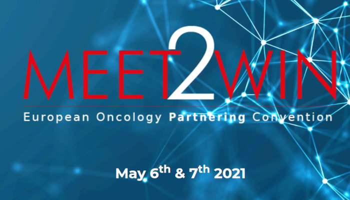
European Oncology Partnering Convention Meet2Win 2021
6 may 2021 -
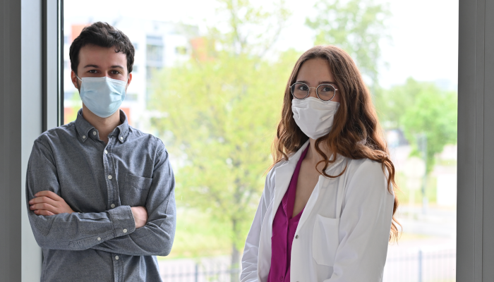
Welcome to our new colleagues
28 apr 2021 -
European project IMLINFO: New article published in Cancers
24 mar 2021 -
Conference paper with IPSEN for Toxins 2021
16 mar 2021 -
Journée thématique sur les innovations en imagerie cutanée
11 mar 2021 -

CROI 2021: Joint work with the Pasteur Institute on HIV-1
6 mar 2021 -
Scientific paper: Mitotic arrest effect on clustering of tumor cells
2 feb 2021 -
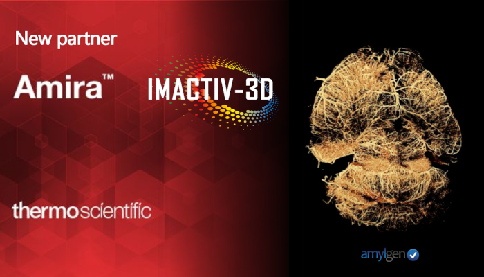
Partnership with ThermoFisher scientific
11 jan 2021 -
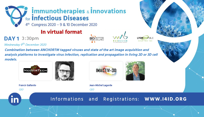
I4ID Congress: Viral infection, replication, propagation and image processing in 2D and 3D
9 dec 2020 -
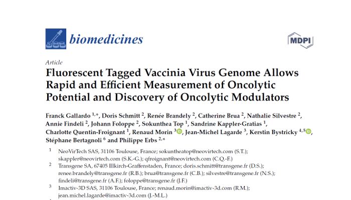
Scientific paper on oncolytic viruses in Biomedicines
4 dec 2020 -
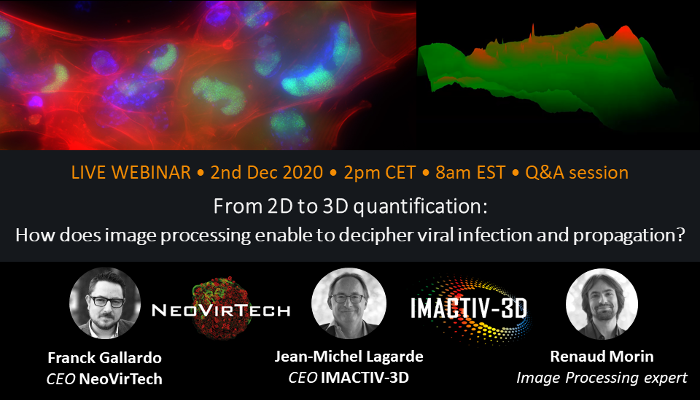
WEBINAR Image processing in 2D and 3D for viral infection and propagation
2 dec 2020 -
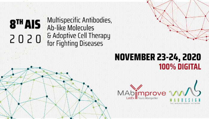
Imactiv-3D at the 8th Antibody Industrial Symposium
23 nov 2020 -
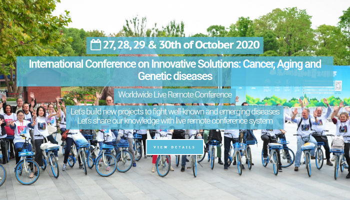
ICIS 2020: Conference on innovative solutions for Cancer, Aging and Genetic diseases
28 oct 2020 -
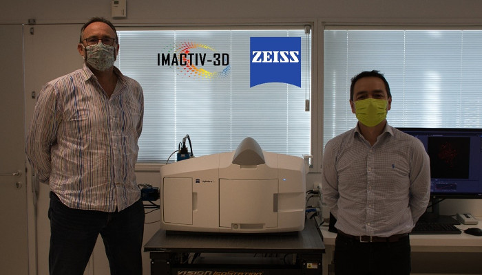
Partnership with Carl Zeiss SAS to offer innovative integrative solution
21 oct 2020 -
Imactiv-3D officially joins the Centre Pierre Potier business incubator
7 jul 2020 -
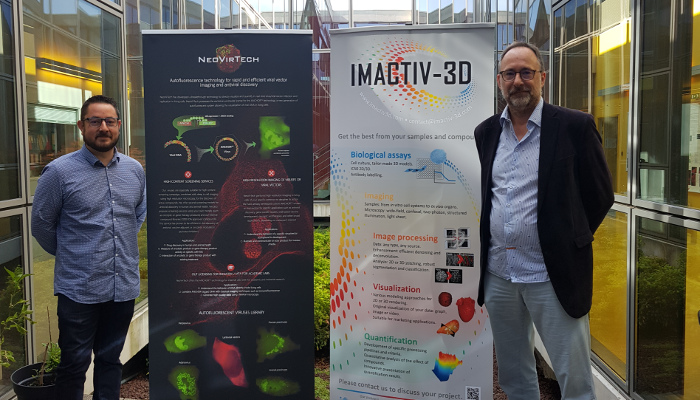
Strategic alliance between Imactiv-3D and NeoVirTech
2 jul 2020 -
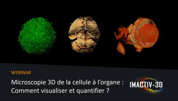
WEBINAR Microscopie 3D de la cellule à l'organe : Comment visualiser et quantifier ?
30 jun 2020 -
Workshop “Mise en lumière des échantillons 3D”
29 apr 2020 -
Ph.D. Defense of Aurélie Gomes
17 dec 2019 -
Start of APELYM 3D project
1 nov 2019 -
Imactiv-3D at Termis workshop on 3D Bioprinting for Cancer
26 aug 2019 -
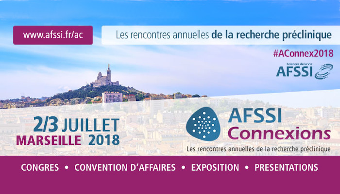
IMACTIV-3D at the AFSSI connexions in Lyon
2 jul 2019 -
AIS Tours : 7th Antibody Industrial Symposium
24 jun 2019 -
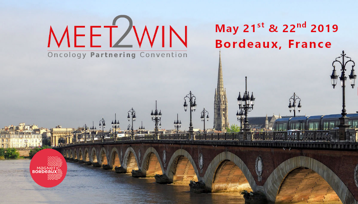
Imactiv-3D at Meet2Win Oncology Partnering Convention
21 may 2019 -
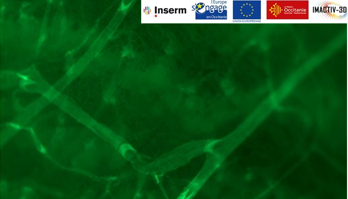
IMALC Project Results
31 dec 2018 -

Joint publication with INSERM in OncoImmunology
17 dec 2018 -

Scientific publication in virology
29 aug 2018 -
Imactiv-3D to attend the AFSSI Connexions
2 jul 2018 -
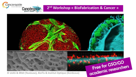
Imactiv-3D Sponsor of the 2nd Workshop BioFabrication & Cancer
27 jun 2018 -
Meet2Win 2018
17 may 2018 -
IMLINFO (EFA281/16): Start of the European project
1 jan 2018 -
Imactiv-3D at the SF2IC Seminar
23 oct 2017 -

Imactiv-3D at the INEXO Symposium
7 jul 2017 -

Start of IMALC Project on Follicular Lymphomas
1 jan 2017 -

Imactiv-3D first scientific publication
12 dec 2016 -
Establishment of the company
13 nov 2015