Imaging
Our unique know-how on a large range of microscopes to get the best out of your biological samples, regardless of their size (sub-cellular, cellular, tissue up to 1 cm3).
- Expert use of microscopes
- Wide-field
- Fluorescence
- Two-photon
- Confocal
- Structured light
- Live videomicroscopy
- Light-sheet imaging
- Consultancy for imaging strategy & modality
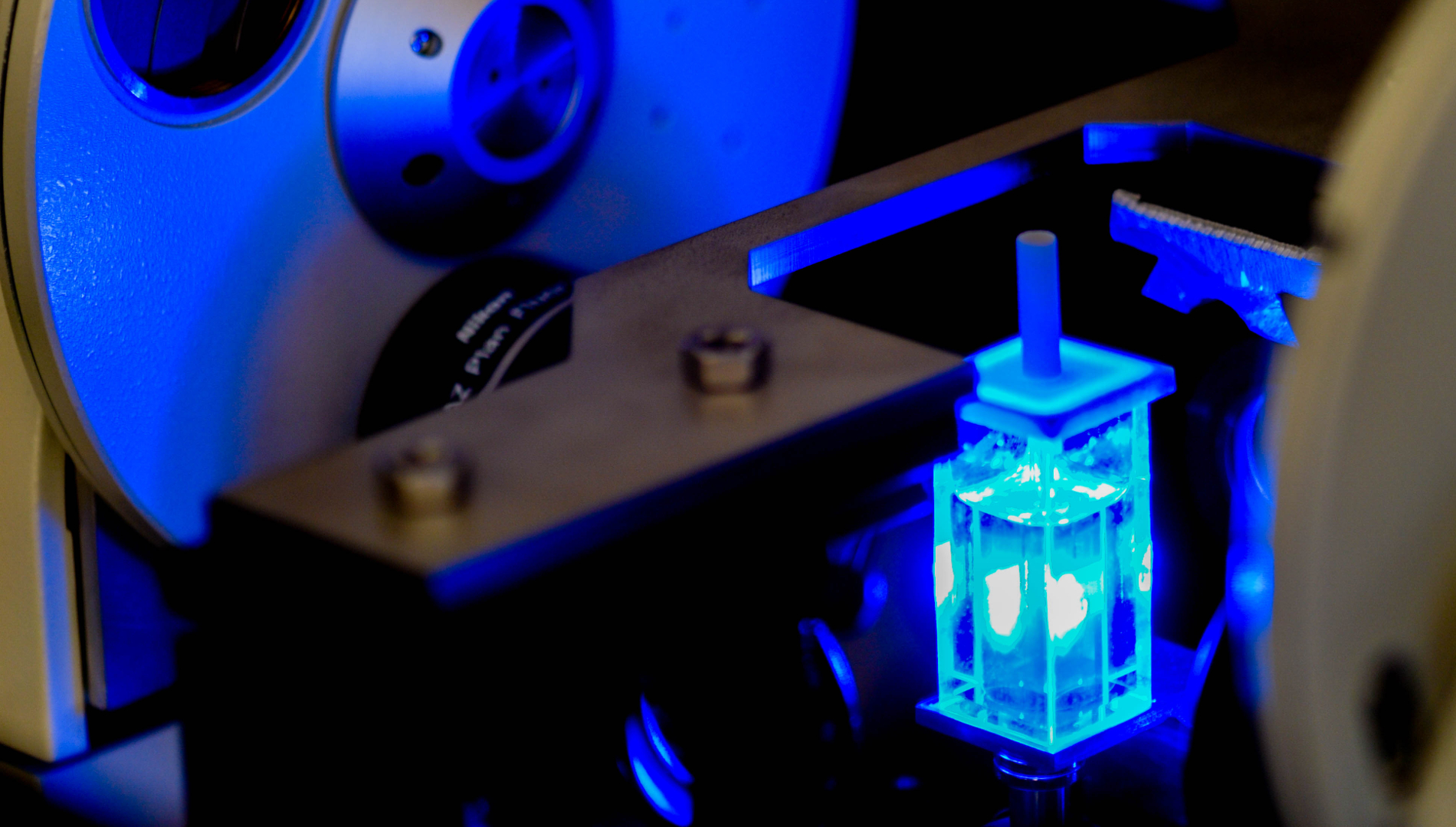
Image Processing

Custom-made algorithms based on state-of-the-art image processing tools and in-house innovation will help you with automating manual processes, improving robustness of your results, reinforcing the reproducibility of your experiments and eventually communicating more efficiently.
- Pre-Processing
- Denoising
- Deblurring
- Image restoration
- Quantification
- Segmentation
- Classification
- Tailor-made algorithms
- Deep learning approaches
- Visualization
- Surface and volume rendering
- 3D animation (with customized scenario)
For more Information
Don't hesitate to contact our team
Sample Preparation
Imactiv-3D brings expertise together to optimize the processing of your biological samples in order to achieve outstanding quality imaging.
- For wide-field microscopy
- Histological sections, cryosections
- Large range of labelling (dyes, antibodies, coloration)
- For 3D imaging
- Sample clearing and inclusion
- In vitro 3D models (spheroids, organoids, bioprinting, RHE…)
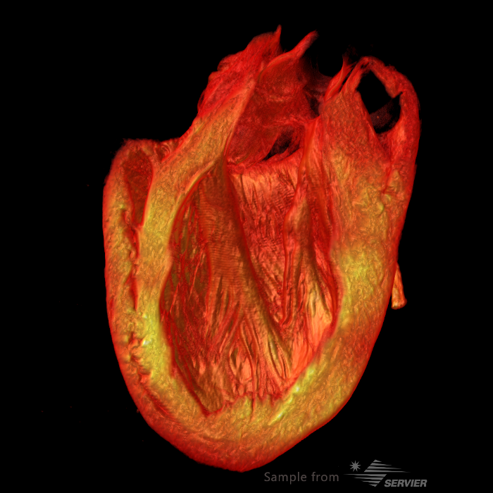
- Ex vivo samples (biopsy, tissue, whole organ)
- Immuno-labelling in toto
Biological Models
Our team of biologists implements cell biology strategy to study your compounds with better in vivo relevance.
- Proliferation & viability analysis
- IC50 on 3D and 2D samples (cytostaticity, cytotoxicity)
- Cell cycle analysis
- 3D cell models
- Free or matrix-embedded spheroids
- Other on demand
- Large range of cell lines
- Cell lines co-culture
- Controlled-atmosphere culture
- Hypoxia
- Physioxia
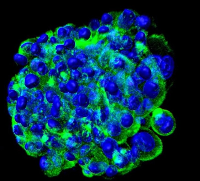
In vitro 3D models that mimic normal or pathological human tissues are essential for understanding the intimate biological mechanisms and for the development of new treatments.
The interest of 3D models is to accurately reproduce the organization of a tissue, recapitulating the cell-cell connections, cell-microenvironment and cellular heterogeneity found in the micro-regions of a tumor or organ. They are attractive models to predictively assess the anti-proliferative activity of new drugs or genotoxic properties.
With their biological relevance and their cost-saving production, in vitro 3D models are a great way to reduce the number of laboratory animals needed, making the research more ethical.