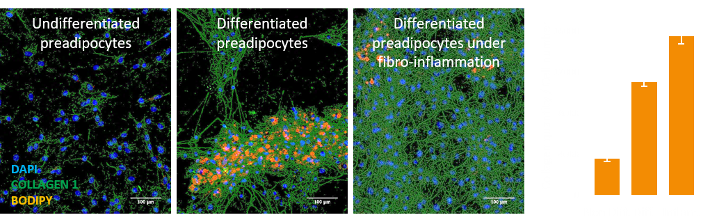
Quantification of fiber production by preadipocytes
Your Needs:
Our Solutions:
- Monitor pre-adipocytes differentiation
- Monitor the fibro-inflammation process (collagen, fibronectin)
Our Solutions:
- Fully automatic imaging and image analysis
- Robust data, short delay and cost-saving

Image acquisition:
- Acquisition done with structured light or confocal microscopy
- Several fields of view to maximize the amount of data
- Image stack for each field of view to get each cellular structure on their focal plane
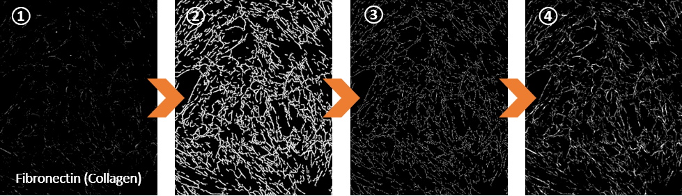
Image processing:
- Detail of an image after acquisition with spinning disk confocal microscopy
- Denoising and segmentation of every fiber
- Refining the segmented object and calculating the criteria: total fiber length, thickness of fibers
- Automatic generation of illustrative images for every field of view
Application example
During weight gain, adipose tissue is characterized by the presence of low-grade inflammation and matrix remodelling. This leads to a greater tissue stiffness and to resistance to weight loss.