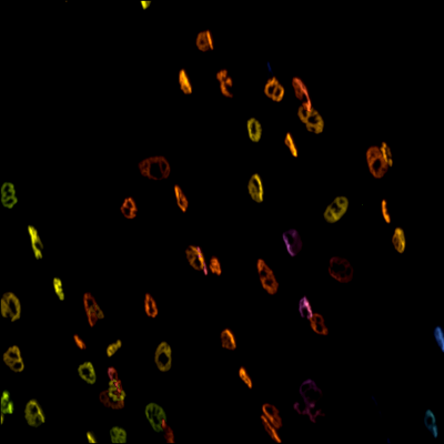
3D ex vivo imaging and characterization of neuromuscular junctions
Your Needs:
Our Solutions:
- Study of neuromuscular pathologies or atrophy models
- Preclinical evaluation of compound efficacy
Our Solutions:
- Light sheet microscopy and clearing to visualize neuromuscular junctions on whole muscle in 3D
- Automated image processing for quantification
General Procedure
Prior to sample collection by Imactiv-3D:
- In vivo labeling by infusion with a fluorescent toxin
- Formalin fixation of extracted sample
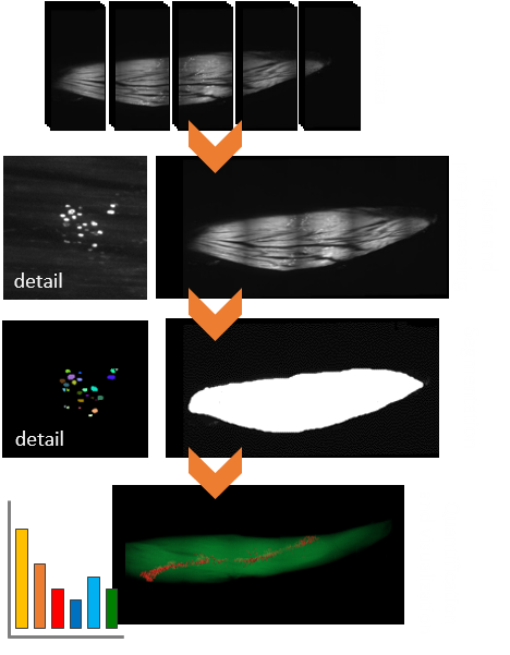
Image acquisition:
- Sample clearing
- 3D light sheet fluorescence microscopy
- Multi-position acquisition
Image processing and analysis:
- Quantitative characterization of all types of leg muscle:
- Estimation of muscle volume
- Absolute number and density of neuromuscular junctions
- 3D visualization with surface and volume rendering:
- Reconstruction of the whole sample
- Visualization of the spatial distribution of junctions
- Advanced display using 3D animations
Application example

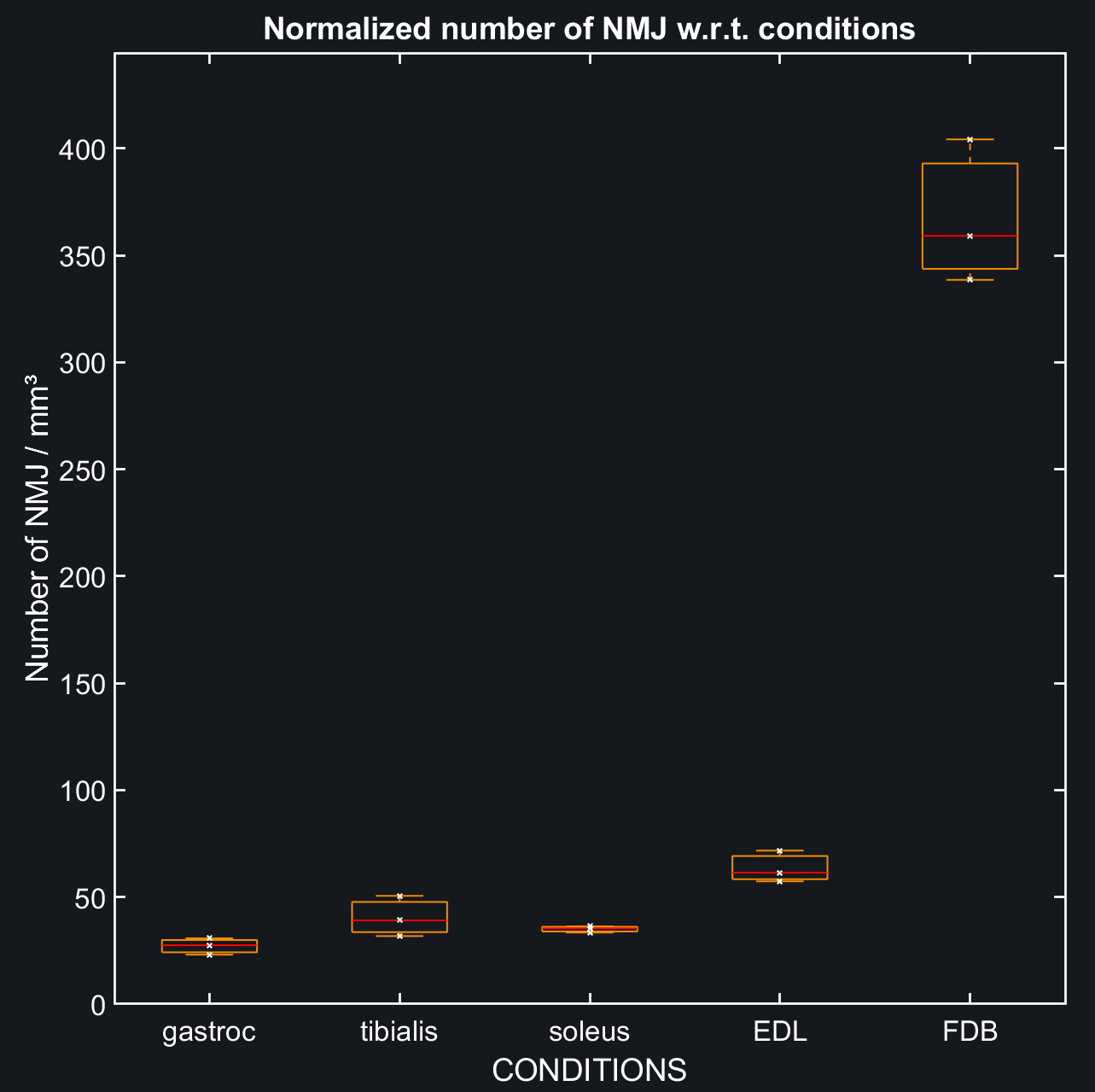
- Aim: comparison of a wide range of muscle types in the rat leg in terms of neuromuscular junction density
- Technical specifications:
- Whole sample acquisition using 3 to 10 fields of view
- Number of junctions ranges from 3 000 to 20 000 depending on the muscle type
- Junction average size is about 60 x 30 x 10 µm³
- Analysis of the neuromuscular junction density following this procedure on 30 muscles produced interesting results among different types of muscle
You can find more informations on our conference paper for Toxicon 2021 available here.