3D imaging of reconstructed skin model
Your Needs:
Our Solutions:
- Qualify 3D models
- Preclinical trials of a compound efficacy on skin structure
Our Solutions:
- Characterize 3D tissue organization over time
- Probes or in toto immunofluorescence labeling
- Light sheet fluorescence microscopy
- Quantification and multiparametric analysis
General Procedure
Prior to sample collection by Imactiv-3D:
- Sample generation and formalin fixation
Tissue processing:
- Possibility to label the sample by probes or in toto immunofluorescence
- Sample clearing
Image acquisition:
- Light sheet microscopy, from 1X to 20X magnification
Image processing and analysis:
- Characterization of cell structure after segmentation: computation of parameters of interest, such as the number of nuclei, the presence of fibers, their morphology, their orientation, the layer thickness…
- 3D visualization with surface and volume rendering

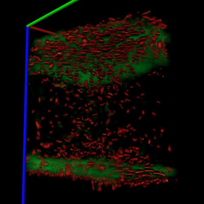
Application example: visualization and monitoring of reconstructed epidermis differentiation.
- Aim:3D visualization of reconstructed epidermis and monitoring of morphological changes in nucleus shape during differentiation.
- Tissue labeling:
- Propidium iodide (red) to monitor nuclei.
- Phalloidin (green) to highlight cortical actin within skin cells.
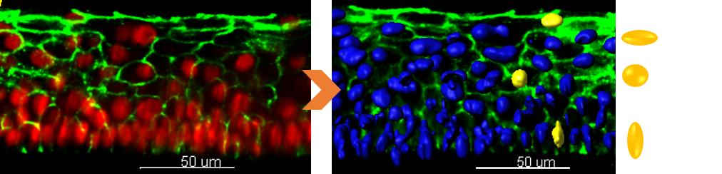
- Images acquired using light sheet fluorescence microscopy.
- Nuclei volume reconstructed in 3D (right figure).