3D ex vivo imaging and characterization of vascular network in rodent
Your Needs:
Our Solutions:
- Study of the impact of pathologies on the cardiac vascular network
- Preclinical study of treatment efficacy
Our Solutions:
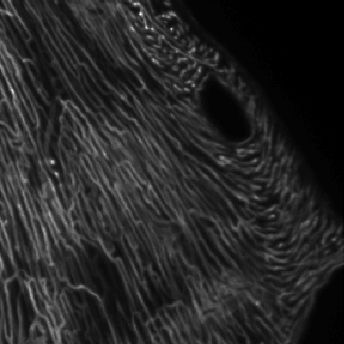
- Light sheet microscopy and clearing to characterize the vascular network in 3D
- Automated 3D image processing for microvascular network quantification
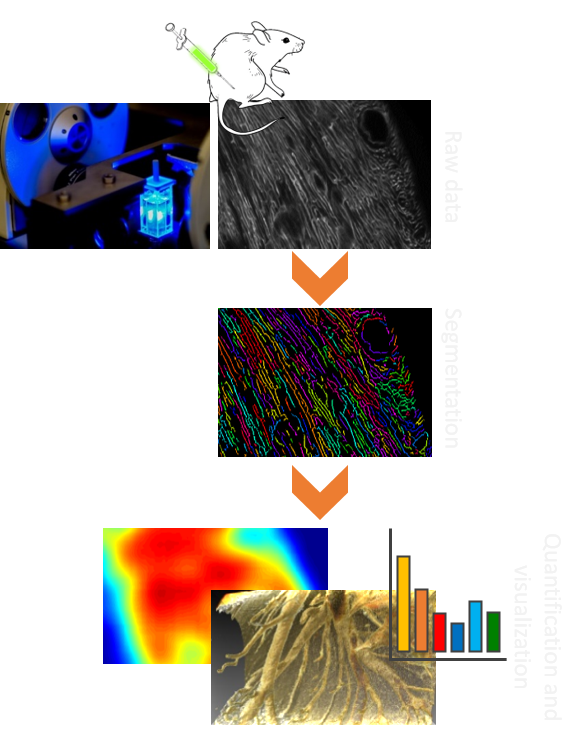
General Procedure
Prior to sample collection by Imactiv-3D:
- In vivo labelling by infusion with a fluorescent lectin before euthanasia.
- Formalin fixation of extracted sample
Image acquisition:
- Sample clearing
- 3D light sheet fluorescence microscopy
- Multi-position acquisition
Image processing and analysis:
- Quantitative characterization of the vascular network
- Vessels segmentation
- Extraction of efficient volume
- Computation of parameters of interest: vessels length and local size, density of the vascular network
- 3D visualization with surface and volume rendering
- Reconstruction of the whole sample
- Advanced display using 3D animations
Application example
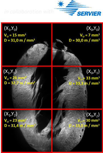
- Aim: Characterization of the microvascular network of a whole rat heart.
- Technical specifications:
- Whole sample acquisition using 6 to 9 fields of view
- Raw data amounts to about 200 GB or 100 billion voxels
- Image processing takes 14 hours using a parallel architecture
- Analysis of the microvascular network in 33 samples following this procedure exhibited an internal variation of only 3.5 % with respect to the mean.
- Assessment of microvascular alteration in pathological rat with comorbidities such as hypertension/diabetes (and effect of a potential treatment on vascular density).