Quantitative morphometric analysis of 3D structures in whole organs
Your Needs:
Our Solutions:
- Study of the morphology of whole organ structures
- Preclinical study of treatment efficacy
Our Solutions:
- Light sheet microscopy and clearing to characterize the vascular network in 3D
- Automated 3D image processing for micro-vascular network quantification
General Procedure
Image acquisition:
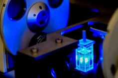
- Sample optical clearing
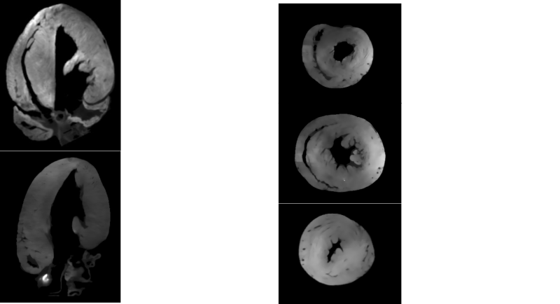
- 3D light sheet auto-fluorescence microscopy (label-free imaging)
- Multi-position acquisition
Image processing and analysis:
- Development of a specific algorithm to characterize 3D structures.
Application example
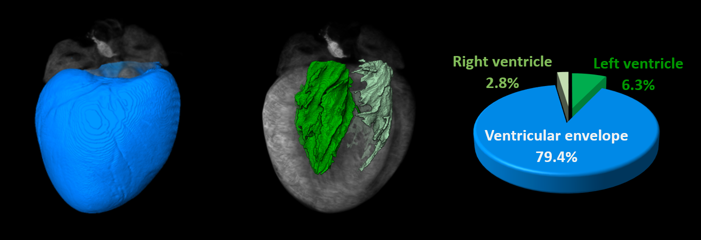
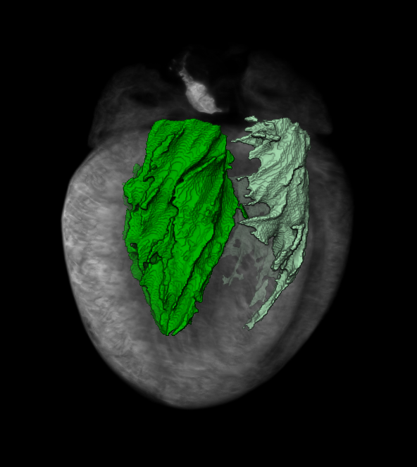
Aim: Analyze of the heart morphology in 3D following the recommendations by the American Heart Association Writing Group on Myocardial Segmentation and Registration for Cardiac Imaging.
Volume rendering and reconstruction of each ventricle allows the quantification of anatomical structures (graphic representation in function of the percentage of total volume).